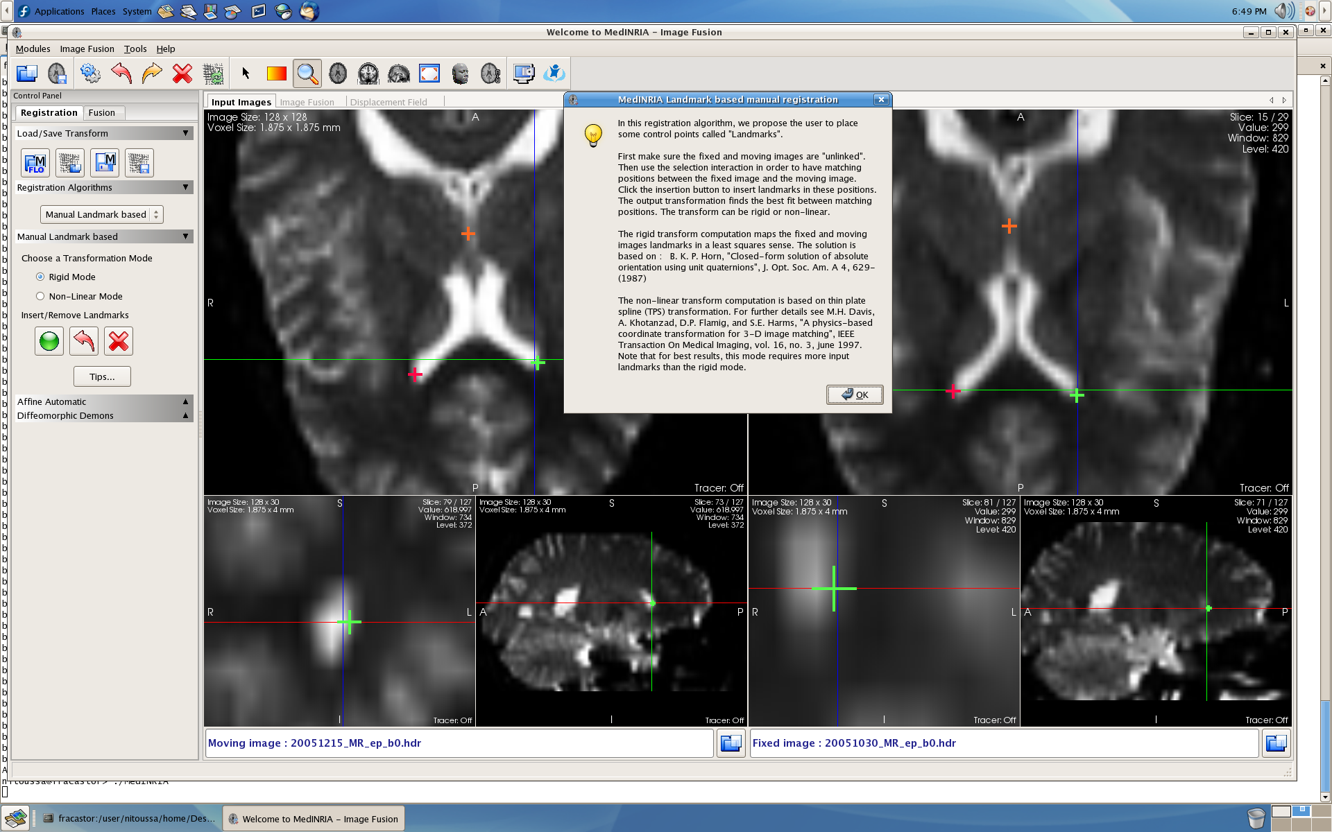

PLoS ONE 11(9):Įditor: Andrea Antal, University Medical Center Goettingen, GERMANY (2016) Investigation of Motor Cortical Plasticity and Corticospinal Tract Diffusion Tensor Imaging in Patients with Parkinsons Disease and Essential Tremor. The microstructure of the CST is not relevant to the deficiency of M1 associative plasticity in PD and ET.Ĭitation: Lu M-K, Chen C-M, Duann J-R, Ziemann U, Chen J-C, Chiou S-M, et al. Findings suggest that both PD and ET with intention tremor have impairment of the associative LTP-like corticospinal excitability change in M1. There was no significant correlation between the PAS effects and the DTI measures. No significant differences of the mean FA and MD were found between groups. SICI and LICI were significantly reduced after PAS irrespective of groups. PAS increased MEP amplitude in HC but not in PD and ET. PAS effects and DTI data were simultaneously examined between groups. The DTI measures of fractional anisotropy (FA) and mean diffusivity (MD) were acquired. The primary motor cortex (M1) excitability, measured by motor evoked potential (MEP) amplitude and by short-interval and long-interval intracortical inhibition (SICI and LICI) was compared between the three groups before and after PAS. Three groups of twelve subjects with moderate severity PD, ET with intention tremor and healthy controls (HC) were studied. Motor circuit dysfunctions can be complementarily investigated by paired associative stimulation (PAS)-induced long-term potentiation (LTP)-like plasticity and diffusion tensor imaging (DTI) of the corticospinal tract (CST). nii file and a result excel sheet.Parkinson’s disease (PD) and essential tremor (ET) are characterized with motor dysfunctions. Each of the extracted image files contains a ROI. The reserve folder contains the following computed images: ADC, FA, L1, L2, 元 and RD along with B0 image. All DWI raw data was converted from bruker format to img/hdr (Analyze) format via MATLAB and stored in individual folder (#_B900). Once unzipped into DTI_data folder, this has two subfolders 'IL' for Males from Illinois and ''KSIL" males from Kansas dams and Illinois males. The processing steps are explained in detail in article.ĭTI Data: The DW-3D-EPI images were processed and analyzed using MATLAB (Mathworks, Natick, MA) and MedINRIA (version 1.9.0 ) software. The filenames of these files is self explanatory. This subject folder contains the EPI Fc-Conn data in NIFTI (.nii) format along with mask and cropped file. Inside these folders there are separate folder for each subject (Vole). Once unzipped into 'FcConnData' folder it has two subfolders for 'IL' for Males from Illinois and ''KSIL" males from Kansas dams and Illinois males.

The article is published in 'Biological Psychiatry: Cognitive Neuroscience and Neuroimaging' with DOI: /10.1016/j.bpsc.2020.08.014įc-Conn Data: The Functional Connectivity data is in "FcConnData.zip" folder. We compared apparent diffusion coefficient (ADC), fractional anisotropy (FA), and BOLD resting-state functional connectivity (rsFC) between males. Behavioral differences between these males are associated with overexpression estrogen receptor alpha in the medial amygdala (MeA) and bed nucleus of the stria-terminalis (BST) and neuropeptide expression in the paraventricular nucleus (PVN).

Specifically, Males from Illinois (IL), who display high levels of prosocial behavior and F1 males from Kansas dams and Illinois males (KI), which display the lowest level of prosocial behavior and higher aggression. We used the highly prosocial prairie vole to test the hypothesis that higher-order brain structure, microarchitecture and functional connectivity, would differ between males from populations with distinctly different levels of prosocial behavior.


 0 kommentar(er)
